Completed by: intern of the Department of Neurology and Neurosurgery, S.A. Valkov. thalamus
Thalamic syndrome is a condition caused by damage to an area of the brain called the thalamic thalamus. The thalamus is a paired formation represented by gray matter and consisting of the anterior tubercle, body and pillow. Refers to the intermediate part of the brain. The nuclei of the optic thalamus are responsible for vision, hearing, tactile sensations, and balance. The thalamus performs the functions of processing information, regulating attention, and coordinating the work of the musculoskeletal system. The part of the brain that coordinates speech, memory, and emotions. Damage to the visual thalamus entails disruption of the described functions.
Main symptoms of thalamic syndrome
The set of symptoms caused by damage to the visual thalamus is otherwise called Dejerine-Roussy syndrome. The painful condition resulting from damage to the thalamus was first described in the 19th century. A detailed definition of the symptoms and causes was given by the French scientists Dejerine and Roussy at the beginning of the 20th century.
The signs of the syndrome are:
- loss of pain and skin sensitivity on one side of the body;
- increased pain perception threshold with the inability to accurately determine its location;
- intense burning pain on one side of the body;
- perversion of sensitivity (temperature stimulus is felt as painful, light touches cause discomfort);
- loss of sensitivity to vibration effects;
- exhaustion and weakening of the muscles of the affected part of the body;
- erratic chaotic movements of the fingers of the upper limb;
- formation of the so-called thalamic hand: the forearm is bent and turned back, the hand is bent, the distal phalanges are straight with the proximal and middle phalanges half-bent;
- unilateral motor coordination disorder;
- partial blindness – lack of perception of the right or left half of the visual field;
- drooping of one corner of the mouth, unilateral facial paralysis;
- impaired concentration.
The patient's psychological state is characterized by mood swings, depression, and suicidal thoughts.
Causes of pathology
Thalamic syndrome is not a disease, but a set of signs and clinical manifestations. The symptom complex can be caused by vascular disorders of the deep branches of the posterior cerebral artery, damage to the ventral posterolateral nucleus of the thalamus. These conditions can lead to:
- injury;
- malignant brain tumor with metastases in the thalamus;
- ischemic stroke;
- hemorrhagic stroke.
The origin of hyperpathic pain and severe psycho-emotional disorders accompanying thalamic syndrome is not fully explained. Other neurological symptoms are caused by the following reasons:
- damage to the structures of the cerebellar dentate-thalamic tract;
- dysfunction of the medial lemniscus;
- damage to the hypothalamic nuclei.

Diagnosis and treatment
Diagnosis is based on a set of measures that involve clinical and instrumental examination methods:
- collecting anamnesis, studying patient complaints and determining possible causes of pathology;
- checking superficial and deep skin sensitivity;
- establishing muscle strength of the limbs;
- visual field check;
- determination of reactions to auditory, visual and taste stimuli;
- computed and magnetic resonance imaging;
- cerebral angiography.
Treatment of the pathology - symptomatic and pathogenetic - is based on the use of antipsychotics and antidepressants. A polypharmacotherapy regimen, a combination of drugs: an anticonvulsant, an antidepressant and an opioid, is considered effective. In cases where conservative methods do not bring results, surgical intervention is indicated, during which the doctor destroys the ventrolateral nucleus of the thalamus. The operation is performed using a minimally invasive stereotactic method.
Along with traditional medicine, treatment of thalamic pain syndrome with folk remedies may be effective. Such therapy is aimed at relieving painful symptoms, but does not affect the causes and mechanisms of pathology.
Traditional medicine suggests treating the syndrome by pain relief or by trying to restore sensitivity to the skin, for which the following recipes can be used.
- Ginger infusion for bathing (to relieve pain): 50 grams of crushed dry root of the plant are placed in a thermos, poured with a liter of boiling water and infused for one hour. The contents are added to the bath. It is necessary to take water procedures for 15 minutes. Daily use of this infusion for bathing is contraindicated. Before taking a bath with ginger for the first time, you need to determine whether there are any allergic reactions to the plant. With a cotton swab moistened with the prepared solution, wipe a small area of skin on the wrist or elbow and wait 15-20 minutes.
- In case of loss of sensitivity, alcohol tincture of dandelions has a healing effect. To prepare it, take 100 grams of the dry matter of the plant and pour half a liter of vodka. Infuse the medicine for a week, leaving the jar in a dark place and periodically shaking the contents. The tincture is used to rub the parts of the body that have lost sensitivity.
Thalamic syndrome is a complex of neurological symptoms caused by damage to the thalamus opticus. Diagnosis of pathology involves the use of clinical and instrumental methods. Treatment is symptomatic and pathogenetic.
Like any other organ of the brain, the thalamus has an extremely important and irreplaceable function for the body. It’s hard to imagine, but this relatively small organ is responsible for all mental functions: perception and understanding, memory and thinking, because thanks to it we see, understand, feel the world and perceive everything that surrounds us. Thanks to its work, we navigate in space and time, feel pain, this “sensitivity collector” perceives and processes information received from all receptors, except the sense of smell, and transmits the necessary signal to the desired part of the cerebral cortex. As a result, the body gives the correct reaction, displays the correct behavior patterns to the corresponding stimulus or signal.
General information
The diencephalon is located under the corpus callosum and consists of: the thalamus (thalamic brain) and the hypothalamus.
The thalamus (aka: visual thalamus, sensitivity collector, body informant) is a section of the diencephalon located in its upper part, above the brain stem. Sensory signals and impulses from various parts of the body and from all receptors (except smell) flow here. Here they are processed, the organ evaluates how important the incoming impulses are for a person and sends the information further to the CNS (central nervous system) or to the cerebral cortex. This painstaking and vital process occurs thanks to the components of the thalamus - 120 multifunctional nuclei that are responsible for receiving signals, impulses and sending processed information to the appropriate one.
Thanks to its complex structure, the “visual thalamus” is capable of not only receiving and processing signals, but also analyzing them.
Ready information about the state of the body and its problems reaches the cerebral cortex, which, in turn, develops a strategy for solving and eliminating the problem, a strategy for further actions and behavior.
Structure
The thalamus is a paired ovoid formation consisting of nerve cells that are united into nuclei, thanks to which the perception and processing of signals and impulses coming from different sense organs occurs. The thalamus occupies the bulk of the diencephalon (approximately 80%). Consists of 120 multifunctional gray matter nuclei. It is shaped like a small chicken egg.
Based on the structure and location of individual parts, the thalamic brain can be divided into: metathalamus, epithalamus and subthalamus.
Metathalamus(subcortical auditory and visual center) - consists of medial and lateral geniculate bodies. The auditory lemniscus ends in the nucleus of the medial geniculate body, and the visual tracts end in the lateral geniculate nucleus.
The medial geniculate bodies constitute the auditory center. In the medial part of the metathalamus, from the subcortical auditory center, cell axons are directed to the cortical end of the auditory analyzer (superior temporal gyrus). Dysfunction of this part of the metathalamus can lead to hearing loss or deafness.
Lateral geniculate bodies constitute the subcortical visual center. This is where the optic tracts end. The axons of the cells form the optic radiation, along which visual impulses reach the cortical end of the visual analyzer (occipital lobe). Dysfunction of this center can lead to vision problems, and severe damage can lead to blindness.
Epithalamus(suprathalamus) - the upper posterior part of the thalamus, which rises above it: includes the pineal gland, which is the supracerebral endocrine gland (pineal gland). The pineal gland is in a suspended state, as it is located on leashes. It is responsible for the production of hormones: during the day it produces the hormone serotonin (the hormone of joy), and at night it produces melatonin (a regulator of the daily routine and the hormone responsible for the color of the skin and eyes). The epithalamus plays a role in the regulation of life cycles, regulates the onset of puberty, sleep and wakefulness patterns, and inhibits the aging process.
Lesions of the epithalamus lead to disruption of life cycles, including insomnia, as well as sexual dysfunction.
Subthalamus(subthalamus) or prethalamus is a small-volume brain substance. It consists mainly of the subthalamic nucleus and has connections with the globus pallidus. The subthalamus controls muscle responses and is responsible for action selection. Damage to the subthalamus leads to motor disturbances, tremors, and paralysis.
In addition to all of the above, the thalamus has connections with the spinal cord, with the hypothalamus, subcortical nuclei and, naturally, with the cerebral cortex.
Each department of this unique organ has a specific function and is responsible for vital processes, without which the normal functioning of the body is impossible.

Functions of the thalamus
The “sensitivity collector” receives, filters, processes, integrates and sends information to the brain that comes from all receptors (except smell). We can say that in its centers the formation of perception, sensation, and understanding occurs, after which the processed information or signal enters the cerebral cortex.
The main functions of the body are:
- processing of information received from all organs (receptors of vision, hearing, taste and touch) senses (except smell);
- managing emotional reactions;
- regulation of involuntary motor activity and muscle tone;
- maintaining a certain level of activity and excitability of the brain, which is necessary for the perception of information, signals, impulses and irritations coming from the outside, from the environment;
- responsible for the intensity and feeling of pain.
As we have already said, each lobe of the thalamus consists of 120 nuclei, which, based on functionality, can be divided into 4 main groups:
- lateral (lateral);
- medial (middle);
- associative.
Reticular group of nuclei (responsible for balance) – responsible for ensuring balance when walking and balance in the body.
The lateral group (vision center) is responsible for visual perception, receives and transmits impulses to the parietal, occipital part of the cerebral cortex - the visual zone.
The medial group (hearing center) is responsible for auditory perception, receives and transmits impulses to the temporal part of the cortex - the auditory zone.
Associative group (tactile sensations) - receives and transmits tactile information to the cerebral cortex, that is, signals emanating from receptors of the skin and mucous membranes: pain, itching, shock, touch, irritation, etc.
Also, from a functional point of view, nuclei can be divided into: specific and nonspecific.
Specific nuclei receive signals from all receptors (except smell). They provide a person’s emotional reaction and are responsible for the occurrence of pain.
Specific kernels, in turn, are:
- external - receive impulses from the corresponding receptors and send information to specific areas of the cortex. Through these impulses feelings and sensations arise;
- internal - do not have direct connections with receptors. They receive information already processed by the relay cores. From them, impulses go to the cerebral cortex to the associative zones. Thanks to these impulses, primitive sensations arise and the relationship between sensory areas and the cerebral cortex is ensured.
Nonspecific nuclei support the general activity of the cerebral cortex, sending nonspecific impulses and stimulating brain activity. Having no direct connection with the cortex, the nonspecific nuclei of the thalamus transmit their signals to subcortical structures.
Separately about the visual thalamus
Previously, it was believed that the thalamus processed only visual impulses, and then the organ received the name - visual thalamus. Now this name is considered obsolete, since the organ processes almost the entire range of afferent systems (except for smell).
The system that provides visual perception is one of the most interesting. The main external organ of vision is the eye, a receptor that has a retina and is equipped with special cells (cones, rods) that transform the light beam and electrical signal. The electrical signal, in turn, passing through the nerve cells enters the lateral center of the thalamus, which sends the processed signal to the central part of the cerebral cortex. Here the final analysis of the signal occurs, thanks to which what is seen is formed, that is, the picture.
What are the dangers of dysfunction of the thalamic zones?
The thalamus has a complex and well-established structure, therefore, if malfunctions or problems arise in the work of even a single zone of the organ, this leads to various consequences, affecting individual functions of the body and even the entire body as a whole.
Before reaching the corresponding center of the cortex, signals from the receptors enter the thalamus, or more precisely, to a certain part of it. If certain nuclei of the thalamus are damaged, then the impulse is not processed, does not reach the cortex, or arrives in an unprocessed form, therefore, the cerebral cortex and the entire body do not receive the necessary information.
Clinical manifestations of thalamic dysfunction depend on the specific affected area and can manifest themselves as: problems with memory, attention, understanding, loss of orientation in space and time, disorders of the motor system, problems with vision, hearing, insomnia, and mental disorders.
One of the manifestations of organ dysfunction may be specific amnesia, which leads to partial memory loss. In this case, the person forgets the events that occurred after damage or injury to the corresponding area of the organ.
Another rare disorder affecting the thalamus is fatal insomnia, which can affect several members of the same family. The disease occurs due to a mutation in the corresponding zone of the thalamus, which is responsible for regulating the processes of sleep and wakefulness. Due to the mutation, the proper functioning of the corresponding area fails, and the person stops sleeping.
The thalamus is also the center of pain sensitivity. When the corresponding nuclei of the thalamus are damaged, unbearable pain occurs or, conversely, complete loss of sensitivity.
The thalamus, and the brain as a whole, continue to remain incompletely studied structures. And further research promises great scientific discoveries and help in understanding this vital and complex organ.
The thalami, or visual thalamus, are located on the sides of the third ventricle and make up up to 80% of the mass of the diencephalon. They are ovoid in shape, with an approximate volume of 3.3 cubic meters. cm and consist of cellular clusters (nuclei) and layers of white matter. Each thalamus has four surfaces: internal, external, superior and inferior. The inner surface of the thalamus forms the lateral wall of the third ventricle. It is separated from the underlying hypothalamus by a shallow hypothalamic groove (sulcus hypothalamics), running from the interventricular foramen to the entrance to the cerebral aqueduct. The inner and upper surfaces are separated by the medullary stripe (stria medullaris thalami). The upper surface of the thalamus, like the inner one, is free. It is covered by the fornix and the corpus callosum, with which it has no fusions. In the anterior part of the superior surface of the thalamus is its anterior tubercle, which is sometimes called the eminence of the anterior nucleus. The posterior end of the thalamus is thickened - this is the so-called thalamic cushion (pulvinar). The outer edge of the upper surface of the thalamus approaches the caudate nucleus, from which it is separated by the border strip (stria terminalis). A vascular groove runs along the upper surface of the thalamus in an oblique direction, which is occupied by the choroid plexus of the lateral ventricle. This groove divides the superior surface of the thalamus into outer and inner parts. The outer part of the upper surface of the thalamus is covered with the so-called attached plate, which makes up the bottom of the central section of the lateral ventricle of the brain. The outer surface of the thalamus is adjacent to the internal capsule, separating it from the lenticular nucleus and the head of the caudate nucleus. Behind the thalamus cushion are the geniculate bodies belonging to the metathalamus. The rest of the lower side of the thalamus is fused with the formations of the hypothalamic region. The thalami are located on the path of the ascending tracts going from the spinal cord and brain stem to the cerebral cortex. They have numerous connections with the subcortical nodes, passing mainly through the loop of the lenticular nucleus (ansa lenticularis). The thalamus consists of cellular clusters (nuclei), delimited from each other by layers of white matter. Each nucleus has its own afferent and efferent connections. Neighboring nuclei form groups. There are: I) anterior nuclei (lis //, anteriores) - have reciprocal connections with the mastoid body and fornix, known as the mastoid-thalamic fascicle (Vic d'Azir fascicle) with the cingulate gyrus, related to the limbic system; 2) posterior nuclei, or nuclei of the tubercle cushion (nucli posteriores) - associated with the associative fields of the parietal and occipital regions; play an important role in the integration of various types of sensory information coming here; 3) dorsal lateral nucleus (nucl. dorsolateral) - receives afferent impulses from the globus pallidus and projects them into the caudal parts of the cingulate gyrus; 4) ventrolateral nuclei (nucli ventrolaterales) - the largest specific nuclei, are the collector of most somatosensory pathways: the medial lemniscus, spinothalamic tracts, trigeminothalamic and gustatory tracts, along which impulses of deep and superficial sensitivity pass, etc.; from here, nerve impulses are sent to the cortical projection somatosensory zone of the cortex (fields 1, 2, 3 and 36, according to Brodmann); 5) medial nuclei (nucli mediates) - associative, receive afferent impulses from the ventral and intralaminar thalamic nuclei, hypothalamus, midbrain nuclei and globus pallidus; efferent pathways from here are sent to the associative areas of the prefrontal cortex, located in front of the motor zone; 6) intralamellar nuclei (intralaminar nuclei, nucll. intralaminares) - constitute the main part of the nonspecific projection system of the thalamus; They receive afferent impulses partly through the ascending fibers of the reticular formation of the nerve trunk, partly through fibers starting from the nuclei of the thalamus. The pathways emanating from these nuclei are sent to the caudate nucleus, putamen, globus pallidus, which belong to the extrapyramidal system, and, probably, to other nuclear complexes of the thalamus, which then send them to the secondary associative zones of the cerebral cortex. An important part of the intralaminar complex is the central nucleus of the thalamus, which represents the thalamic section of the ascending reticular activating system. The thalami are a kind of collector of sensory pathways, a place in which all the pathways conducting sensory impulses coming from the opposite half of the body are concentrated. In addition, olfactory impulses enter its anterior nucleus through the mastoid-thalamic fascicle; taste fibers (axons of second neurons located in the solitary nucleus) end in one of the nuclei of the ventrolateral group. Thalamic nuclei that receive impulses from strictly defined areas of the body and transmit these impulses to the corresponding limited zones of the cortex (primary projection zones) are called projection, specific or switching nuclei. These include the ventrolateral nuclei. The switching nuclei for visual and auditory impulses are located, respectively, in the lateral and medial geniculate bodies, adjacent to the posterior surface of the visual tuberosities and constituting the bulk of the abdomen. The presence in the projection nuclei of the thalamus, primarily in the ventrolateral nuclei, of a certain somatotopic representation makes it possible, with a pathological focus in the thalamus limited in volume, to develop a sensory disorder and associated motor disorders in any limited part of the opposite half of the body. Associative nuclei, receiving sensitive impulses from switching nuclei, subject them to partial generalization - synthesis; as a result, impulses are sent from these thalamic nuclei to the cerebral cortex, already complicated due to the synthesis of information arriving here. Consequently, the thalamus is not only an intermediate switching center, but can also be a place for partial processing of sensitive impulses. In addition to the switching and associative nuclei, the thalamus contains, as already mentioned, intralaminar (parafascicular, median and medial, central, paracentral nuclei) // reticular nuclei, which do not have a specific function. They are considered as part of the reticular formation and are united under the name of the nonspecific diffuse thalamic system. Being associated with the cerebral cortex and the structures of the limbic-reticular complex. This system takes part in the regulation of tone and in the “tuning” of the cortex and plays a certain role in the complex mechanism of the formation of emotions and the corresponding expressive involuntary movements, facial expressions, crying and laughter. Thus, information from almost all receptor zones converges to the thalami along afferent pathways. This information is subject to significant processing. From here, only part of it is directed to the cerebral cortex, while the other and probably the majority part takes part in the formation of unconditioned and, possibly, some conditioned reflexes, the arcs of which close at the level of the thalamus and formations of the striopallidal system. The thalami are the most important link in the afferent part of the reflex arcs, which determine instinctive and automated motor acts, in particular habitual locomotor movements (walking, running, swimming, cycling, skating, etc.). Fibers going from the thalamus to the cerebral cortex take part in the formation of the posterior femur of the internal capsule and the corona radiata and form the so-called radiation of the thalamus - anterior, middle (upper) and posterior. The anterior radiate connects the anterior and partly the internal and external nuclei with the cortex of the frontal lobe. The middle radiation of the thalamus - the widest - connects the ventrolateral and medial nuclei with the posterior parts of the frontal lobe, with the parietal and temporal lobes of the brain. The posterior radiation consists mainly of visual fibers (radiatio optica, or Graziole's bundle), going from the subcortical visual centers in the occipital lobe, to the cortical end of the visual analyzer, located in the area of the calcarine sulcus (fissura calcarina). The corona radiata also contains fibers that carry impulses from the cerebral cortex to the thalamus (corticothalamic connections). The complexity of the organization and variety of functions of the thalamus determines the polymorphism of possible clinical manifestations of its damage. Damage to the ventrolateral part of the thalamus usually leads to an increase in the sensitivity threshold on the side opposite to the pathological focus, while the affective coloring of pain and temperature sensations changes. The patient perceives them as difficult to localize, diffuse, and having an unpleasant, burning tint. Characteristic in the corresponding part of the opposite half of the body is hypalgesia in combination with hyperpathy, with a particularly pronounced disorder of deep sensitivity, which can lead to awkwardness of movements and sensory ataxia. With damage to the posterolateral part of the thalamus, the so-called thalamic Dejerine-Roussy syndrome may appear (described in 1906 by French neurologists J. Dejerine (1849-1917) and G. Roussy (1874-1948)1, which includes burning, painful, sometimes unbearable thalamintic pain in the opposite half of the body in combination with a violation of superficial and especially deep sensitivity, pseudoasteriognosis and sensitive hemiataxia, symptoms of hyperpathia and dysesthesia. Thalamic Dejerine-Roussy syndrome most often occurs when an infarction focus develops in it connection with the development of ischemia in the lateral arteries of the thalamus (aa. thalamki iaterales) - branches of the posterior cerebral artery. Sometimes, on the side opposite the pathological focus, transient hemiparesis occurs and homonymous hemianopsia develops. Sensitive hemiataxia can be a consequence of a disorder of deep sensitivity , pseudoastriognosis. In case of damage to the medial part of the thalamus, the dentate-thalamic pathway along which impulses from the cerebellum pass to the thalamus, and rubrothalamic connections on the side opposite to the pathological focus, ataxia appears in combination with athetoid or choreoathetoid hyperkinesis, usually especially pronounced in hands and fingers (“thalamic” hand). In such cases, there is a tendency to fix the hand in a certain position: the shoulder is pressed to the body, the forearm and hand are bent and pronated, the main phalanges of the fingers are bent, the rest are extended. At the same time, the fingers make slow, elaborate movements of an athetoid nature. The arterial blood supply to the thalamus includes the posterior cerebral artery, posterior communicating artery, anterior and posterior villous arteries.
PERFORMED BY: INTERN OF THE DEPARTMENT OF NEUROLOGY AND NEUROSURGERY SSMU VALKOV S.A. THALAMUS. ANATOMY, FUNCTIONS AND SYMPTOMS OF THE LESION Presentations
ANATOMY OF THE THALAMUS A large accumulation of gray matter formed by several groups of nuclei Occupies the central part of the brain Has numerous afferent and efferent connections with its various parts Presentations


ANATOMY. NUCLEI OF THE THALAMUS Each thalamus consists of several groups of nuclei separated by the inner medulla. In the anterior sections, three large nuclear groups are distinguished: 1) anterior 2) medial 3) lateral, divided into ventral and dorsal parts

ANATOMY. NUCLEI OF THE THALAMUS Between the medial and lateral nuclei in the area of the internal medullary plate there are several nuclei, small intralaminar nuclei and the centromedial nucleus. The posterior parts of the thalamus are represented by a large nuclear mass, called the “thalamic cushion”. In the ventral part, the medial and lateral geniculate bodies are adjacent to the pillow

ANATOMY OF THE THALAMUS The outer medulla separates the thalamus from the reticular nucleus, which consists of an arcuate cord of cells located on the border between the lateral thalamic nucleus and the internal capsule. The posterior sections of the internal capsule delimit the thalamus from the globus pallidus


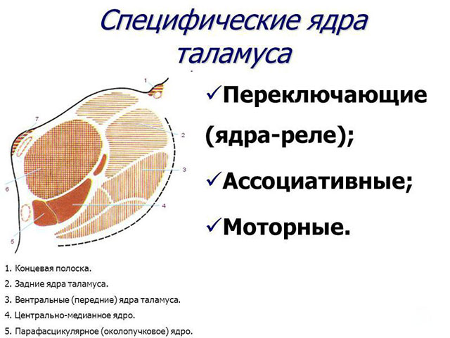
SPECIFIC SENSORY NUCLEI OF THE THALAMUS The ventral group of nuclei, the ventral posterolateral and ventral posteromedial, receive afferentation from the sensory conductors of the spinothalamic tract, the medial lemniscus and the trigeminal lemniscus and project it onto the somatosensory cortex of the posterior central gyrus. The nuclei of the external geniculate bodies receive information from the visual tracts and project it to the visual cortex of the occipital lobe. Nuclei of the internal geniculate bodies - receive afferentation from the auditory pathways on both sides and project it to the auditory cortex of the temporal lobe

SPECIFIC MOTOR NUCLEI OF THE THALAMUS Anterior ventral nucleus - receives afferentation from the globus pallidus Ventrolateral nucleus - receives afferentation from the globus pallidus and the contralateral dentate nucleus of the cerebellum. They transmit information from the cerebellum and globus pallidus to the motor cortex of the precentral gyrus in a strict somatotopic order, participating in the formation of coordinated precise goal-directed movements

NON-SPECIFIC NUCLEI OF THE THALAMUS Non-specific nuclei are not associated with any one sensory or motor modality. Efferent impulses are sent to the caudate nucleus, putamen, globus pallidus and other thalamic nuclei, which transmit them to the associative zones of the cortex. Nonspecific nuclei of the thalamus are part of the activating reticular formation of the brainstem

NON-SPECIFIC NUCLEI OF THE THALAMUS Intralamnar nuclei - located within the internal medulla. They are a rostral continuation of the reticular formation of the midbrain. The projections of these nuclei are widely represented in the cortex of various lobes of the brain and subcortical nuclei.

INTRALAMMARNAL NUCLEI The largest among them is the central median nucleus. The cells of this nucleus receive afferent information via ascending fibers from the reticular formation of the brainstem and from the cortical nucleus of the cerebellum, as well as from the internal segment of the globus pallidus and some other nuclei of the thalamus. Next, the axons of the cells of the central median nucleus are directed not to the cerebral cortex, but to the caudate nucleus, putamen and globus pallidus. The central median nucleus is an important part of the intralamellar cell complex, forming the thalamic part in the ascending activating system of the reticular formation

NON-SPECIFIC NUCLEI OF THE THALAMUS The reticular nucleus has the shape of a shield, located lateral to the thalamus, and is separated from it by the outer medulla. All thalamocortical projections pass through the reticular nucleus, giving it collateral branches, through which the activities of various thalamic nuclei and neurons of the projection zones are coordinated. Connections between the reticular nucleus and the ascending reticular formation provide extensive interaction between the thalamic nuclei and the activating and inhibitory systems of the brain.


ASSOCIATIVE NUCLEI OF THE THALAMUS The dorsolateral nucleus is connected with the cingulate cortex and is involved in memory processes. The dorsomedial nucleus receives afferentation from the globus pallidus, olfactory tract, and amygdala and is connected to the prefrontal cortex. It is involved in the processes of cognition, the formation of judgments and mood. The thalamic cushion receives afferentation from the primary visual centers of the superior colliculus and lateral geniculate body and projects it to the associative visual and parietal cortex. These connections, not being an independent source of conscious visual perception, form the function of attracting attention to objects of interest located in the peripheral visual field.

PUNCHES OF THE THALAMUS Connections between the thalamus and the cerebral cortex pass through four thalamic peduncles, united in the corona radiata. The anterior thalamic peduncle passes through the anterior femur of the internal capsule, reaching the prefrontal cortex and cingulate gyrus. The superior thalamic peduncle passes through the posterior femur of the internal capsule and is directed to the motor and sensory projections of the precentral and postcentral gyri. The posterior thalamic peduncle, through the posterior femur of the internal capsule, reaches the cortical projections of the occipital lobe, as well as the posterior cortex of the parietal and temporal lobes. The inferior thalamic peduncle goes to the anterior parts of the temporal lobe and orbital cortex.

PHYSIOLOGY OF THE THALAMUS The functions of the thalamus are varied and complex, which is due to its extensive connections both with the periphery and with all parts of the nervous system. The thalamus is the relay station for all sensory information, both exteroceptive and interoceptive. By transmitting it to the cerebral cortex, it becomes conscious and is used in the formation of complex motor and mental functions, including consciousness.


PHYSIOLOGY OF THE THALAMUS Bilateral connections of the thalamus with subcortical formations and the hypothalamus play a role in processing sensory information and giving it a certain emotional expressiveness. The feeling of pain or pleasure is manifested by specific emotional and autonomic reactions: facial movements, changes in the size of the pupils, redness or paleness of the skin, crying, increased breathing and heart rate, independent of consciousness


BLOOD SUPPLY OF THE THALAMUS The blood supply of the thalamus comes from the system of vertebrobasilar arteries, partially with branches from the posterior communicating artery of the carotid basin. The thalamotuberous, or polar, artery, arising from the posterior communicating artery, supplies the anterior parts of the thalamus. Thalamoperforating, or paramedian, arteries depart from the PCA in the area of the bifurcation of the main or from the posterior communicating artery. They supply blood to the dorsomedial sections, intralaminar nuclei, and mamillothalamic tract. Thalamocenecular arteries, the number of which reaches 5-6, arise from the PCA and supply blood to the ventrolateral sections of the thalamus. The posterior villous arteries (external and internal) are directed to the cushion and the internal and external geniculate bodies. Vertebrobasilar basin: 1 main artery; 2 ZMA; 3 thalamo-perforating (paramedian) arteries; 4 thalamogeniculate arteries; 5 posterior villous arteries. Carotid basin: 6th internal carotid artery; 7 posterior communicating artery; 8 polar (thalamotuberous) arteries; 9 anterior villous arteries


THALAMOTUS ARTERY SYNDROME Since the anterior parts of the thalamus are projected onto the prefrontal cortex of the frontal lobes, their damage resembles the clinic of frontal syndrome: the patient experiences apathy and abulia, he is uninitiative and sloppy. Bilateral damage to the poles of both visual thalamus leads to severe neuropsychological disorder, manifested by aspontaneity and severe amnestic disorders; these changes are persistent and do not regress

INFARCTS IN THE ZONE OF THALAMOPERFORANT ARTERIES Manifest by acute depression of consciousness due to damage to the intralaminar nuclei and reticular formation of the midbrain. These disorders may present as hypersomnia, where the patient can be awakened, but when the stimulation is stopped, he or she falls back into deep sleep or coma. Another characteristic sign is a violation of vertical gaze, most often paresis of upward gaze, but it can also be downward, very rarely convergent strabismus. Possible hyperkinesis: asterexis, rubral tremor, dystonia, blepharospasm

INFARCTS IN THE BASIS OF THE THALAMOCENIC ARTERY In these cases, Dejerine Roussy syndrome develops, which is manifested by the occurrence contralateral to the lesion: Hemihypesthesia, including a disorder of all types of sensitivity, mostly deep, often spontaneous paresthesia; Spontaneous pain with hyperpathic coloring, treatment of which with non-narcotic analgesics is ineffective; Sensitive hemiataxia; Astereognosis; Homonymous hemianopsia; Hemiparesis, persistent or transient (persistent or temporary damage to the adjacent internal capsule); Specific installation of the hand “thalamic” hand with pseudoathetosis movements; Emotive paresis of the facial muscles of the opposite half of the face (absence of involuntary movements accompanying emotions, with complete preservation of voluntary movements), Affective disorders in the form of violent crying or laughter

INFARCTIONS IN THE BASIS OF THE THALAMICENIC ARTERY Damage to this vascular basin may be characterized by a sensory thalamic stroke, a violation of all types of sensitivity according to the hemitype without any additional symptoms. Hemihypesthesia, as a rule, is mild and incomplete, since part of the spinal sensory fibers (spinoreticular tract) bypasses the thalamus. In the future, against the background of regression of disorders, the development of thalamic pain syndrome is possible

INFARCTS IN THE BASIS OF THE THALAMOCENIC ARTERY Sensorimotor thalamic stroke occurs when the focus of ischemia spreads to the posterior thigh of the internal capsule adjacent to the ventrolateral parts of the thalamus. The result is a combination of hemihypesthesia and spastic hemiparesis with increased tendon reflexes and the presence of extensor pyramidal signs (Babinski's symptom)

STROKE IN THE POSTERIOR VILLUS ARTERIES BASIS Manifested by visual impairment (superior quadrant or inferior quadrant hemianopsia, sectoral vision loss). These defects are formed due to damage to the lateral geniculate body. Possible mild sensory and motor disorders (hemiparesis, hemihypesthesia) and neuropsychological disorders - apathy, elements of aphasia, visual hallucinations, etc.

THROMBOSIS OF THE PROXIMAL DIVISION OF THE PCA Leads to damage to the occipital and temporal lobes of the cerebral hemispheres and simultaneous damage to the thalamus as a result of blockage of the thalamogenicular arteries; at the same time, the paramedian arteries of the midbrain can also be affected. As a result, the patient develops homonymous complete or quadrant hemianopsia with preservation of macular vision, sensory aphasia, amnestic disorders, as well as signs of damage to the external parts of the thalamus, hemihypesthesia of all types of sensitivity, Dejerine Roussy syndrome


7.3. Thalamus
The brainstem ends in the very anterior parts with a cluster of gray matter, already divided in half by the third ventricle of the brain into the rudiments of the left and right hemispheres.
In the lower stages of phylogeny, before the appearance of the real brain, these large basal ganglia were the terminal station for all sensory impulses; They also contain mechanisms for the implementation of motor reactions of the body. The focus of the sensitive subcortical centers is the thalamus (Fig. 19, 20). The thalamus develops from the lateral wall of the diencephalon ( diencephalon) in the area of protrusion of the optic vesicles and therefore was previously called the optic thalamus ( thalamus opticus).
Rice. 19. A sagittal section at the level of the diencephalon and brainstem demonstrates the junction of the diencephalon and midbrain, as well as the structures surrounding the third ventricle (according to P. Duus):
1 – pineal gland; 2 – posterior commissure; 3 – leash core; 4 – medullary stripes of the thalamus; 5 6 – pedicle of the arch; 7 – transparent partition; 8 – interventricular foramen (Monroe); 9 – anterior commissure; 10 – interthalamic fusion; 11 – hypothalamic sulcus; 12 – end plate; 13 – visual deepening; 14 – optic chiasm (chiasma); 15 – deepening of the funnel; 16 – pituitary gland; 17 – neurohypophysis; 18 – gray tubercle; 19 – mastoid body; 20 – quadrigeminal plate; 21 – midbrain aqueduct; 22 – IV ventricle

Rice. 20. Section of the diencephalon in the frontal plane (according to P. Duus):
1 – vascular basis of the third ventricle; 2 – hypothalamus; 3 – nucleus of the mastoid body; 4 – subthalamic nucleus; 5 – corpus callosum; 6 – choroid plexus of the third ventricle; 7 – vault; 8 – choroid plexus of the lateral ventricle; 9 – body of the caudate nucleus; 10 – zonal layer; 11 – reticular nucleus of the thalamus; 12 – internal and external medullary plates of the thalamus; 13 – thalamus, lateral group of nuclei; 14 – thalamus, centromedian nucleus; 15 – thalamus, medial group of nuclei; 16 – III ventricle, interthalamic fusion; 17 – pale ball; 18 – internal capsule; 19 – visual tract; 20 – undefined zone; 21 – mastoid-thalamic tract; 22 - cerebral peduncle
Gray matter, which is part of the thalamus, forms several groups of thalamic nuclei: anterior (anterodorsal, anterioventral, anteromedial); median (anterior and posterior paraventricular nuclei, rhomboid nucleus and connecting nucleus); medial (dorsal medial nucleus); ventral (dorsal nucleus, anterior ventral nucleus, ventrolateral nucleus, posterolateral ventral nucleus, posteromedial ventral nucleus, centromedian nucleus, posterolateral nucleus); posterior group (nucleus of the lateral geniculate body, nucleus of the medial geniculate body, cushion nuclei) (Fig. 20, 21). There are a total of 150 thalamic nuclei.
Functionally, three main groups of nuclei can be distinguished:
1. A complex of specific, or relay, thalamic nuclei through which afferent impulses of a certain modality are conducted (tactile, pain, visual, auditory impulses, etc.); These are mainly the anterior parts of the thalamus, the lateral and medial geniculate bodies, and the leash.
2. Nonspecific thalamic nuclei, through which afferent impulses of uncertain modality pass with their diffuse projection in the cerebral cortex (visceral sensitivity, proprio- and enteroreception with switching to vasomotor reactions, intrasecretory and other processes), mainly the medial parts of the tubercle, periplastular parts ( partes paralaminares) and reticular nucleus ( nucl. reticularis thalami), as well as the subthalamic nucleus.

Rice. 21. Nuclei of the thalamus (according to P. Duus):
1 – anterior nuclei of the thalamus; 2 – anteroventral nucleus; 3 – anterior ventrolateral nucleus; 4 – ventral intermedial nucleus; 5 – posterior ventrolateral nucleus; 6 – posteromedial ventral nucleus; 7 – dorsal lateral nucleus; 8 – posterior lateral nucleus; 9 – intralamellar (intralaminar) nuclei; 10 – dorsal medial nucleus; 11 – centromedian nucleus; 12 – thalamic cushion; 13 – medial geniculate body; 14 – lateral geniculate body
3. Associative nuclei of the thalamus, through which impulses pass from other nuclei of the thalamus to the associative (secondary and tertiary) fields of the cerebral cortex; These are mainly the lateral parts of the thalamus, the pillow ( pulvinar thalami).
The morphofunctional study of the thalamus is far from complete, so there are different options for combining subcortical formations. Yes, leash ( habenula) belongs to the epithalamus (together with the pineal gland and posterior commissure); geniculate bodies - to the metathalamus; subthalamic nucleus - to the hypothalamus. Currently, cortical-subcortical relationships involving the thalamus are represented as a vicious circle: thalamus - frontal lobes - caudate nucleus - pallidum - thalamus, etc.
Clinical manifestations of thalamic lesions are characterized by the following triad of signs: hemianesthesia, hemiataxia, hemianopsia.
Total contralateral hemianesthesia (or hemihypesthesia) of thalamic origin is often combined with hemialgia: painful sensations of either burning or coldness. A drop of cold water can cause extremely excruciating pain. Simply touching the skin is extremely painful. Any perceptions (visual, auditory, olfactory) can cause hyperalgesia due to a decrease in the threshold of irritation. Negative emotions worsen the patient’s condition, positive emotions improve it, with the formation of an addiction to pleasant affects.
Hemiataxia thalamic origin is of a mixed sensitive-cerebellar nature.
Homonymous contralateral hemianopsia – loss of the visual fields of the same name (right or left) opposite to the lesion, is a consequence of damage to the lateral geniculate bodies and the pillow.
Other symptoms of damage to the thalamus include the phenomenon of “emotional facialis,” i.e. paresis of the facial muscles (especially weak mobility of the corner of the mouth) on the side opposite to the affected thalamus in a patient with emotional facial expressions (smile) while maintaining the performance of facial movements on task. Also, lesions of the thalamus are characterized by peculiar contractures or postures, for example, “obstetrician’s hand”: the hand is bent at the wrist joint and brought to the ulnar side, straight fingers are brought to the center “in a duck”, the forearm can be bent and pronated. When trying to straighten the arm and stretch it forward, severe ataxia and choreic athetosis (thalamostriatal connections) appear; the rigidity characteristic of capsular contractures is absent.
Other syndromes involving the thalamus
Dejerine–Roussy syndrome: hemianesthesia, hemiataxia, hemihyperpathy with paroxysmal pain, mild transient hemiplegia without contractures, choreiform and athetotic movements, forced laughter, crying.
Foix–Chiari–Nicolescu syndrome, or superior red nucleus syndrome: hemitremor of an intentional nature, mild sensory disorders, unstable choreoathetosis, sometimes in combination with a thalamic arm (unfixed Gilman contracture).
| |


